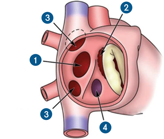
An atrial septal defect is an opening in the atrial septum, or dividing wall between the two upper chambers of the heart, known as the right and left atria.

ASD can be a congenital (present at birth) heart defect, or it can result from the failure of normal postnatal closure of a hole that is present in the heart of every fetus.

An atrial septal defect allows oxygenated blood to pass from the left atrium, through the opening in the septum, and then mix with de oxygenated blood in the right atrium.

1. Ostium secundum atrial septal defect
This is the most common atrial septal defect, affecting over two-thirds of people with atrial septal defects. It is caused when a part of the atrial septum fails to close completely while the heart is developing. This causes an opening to develop in the center of the wall separating the two atria.
2.Ostium primum atrial septal defect.
This defect is part of atrioventricular canal defects and is associated with a split (cleft) in one of the leaflets of the mitral valve.
3. Sinus venosus atrial septal defect.
This defect occurs at the superior vena cava and right atrium junction in the area where the right pulmonary veins enter the heart. As a result, the drainage of one or more of the pulmonary veins may be abnormal in that the pulmonary veins drain to the right atrium, rather than the left atrium.
4. Coronary sinus atrial septal defect.
This defect is located within the wall of the coronary sinus, where it passes behind the left atrium. The coronary sinus carries the blood flow from the heart's own vein, into the right atrium. It is the rarest of all atrial septal defects.